Hydatid Liver Cyst
Understanding Hydatid Disease and Modern Treatment Options
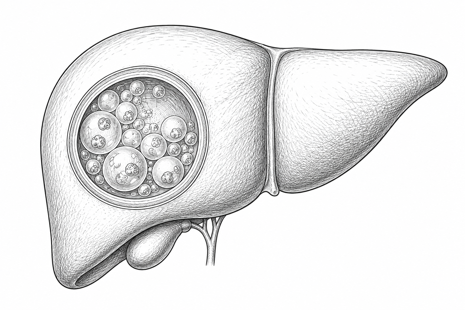
A hydatid liver cyst is a parasitic infection that most commonly affects the liver. Although it can become large and cause complications, modern medical and surgical treatments are highly effective when performed by experienced specialists.
Overview
What Is a Hydatid Liver Cyst?
A hydatid cyst is caused by infection with the parasite Echinococcus granulosus.
After entering the body, the parasite may travel to the liver and slowly form a fluid-filled cyst.
How It Spreads
How Does Hydatid Disease Spread?
Dog (Definitive Host)
The dog carries the parasite in its intestines and sheds eggs in faeces
Parasite Eggs Released
Eggs are shed into the surrounding environment
Contaminated Food or Water
Eggs contaminate soil, vegetables, and water sources
Humans Accidentally Swallow Eggs
Through contaminated food, water, or contact with dogs
Parasite Reaches the Liver
The parasite travels via the bloodstream to settle in the liver
Hydatid Cyst Develops
The cyst grows slowly over months or years
Hydatid disease cannot be transmitted directly from one person to another.
Endemic Regions
Where Is Hydatid Disease Common?
Hydatid disease is more common in rural farming communities where dogs and livestock live in close proximity. Most affected regions include:
Symptoms
Symptoms of Hydatid Liver Cyst
Many hydatid cysts cause no symptoms and are discovered incidentally.
Large cysts may remain silent for many years before causing symptoms.
Complications
Possible Complications
Early specialist assessment significantly reduces the risk of these complications.
Infection
The cyst may become infected with bacteria, causing fever and severe pain.
Rupture
The cyst may rupture into the abdominal cavity, causing serious complications.
Bile Duct Communication
The cyst may open into the bile ducts, leading to jaundice or cholangitis.
Allergic Reaction
Leakage of cyst contents may cause allergic reactions, including rare anaphylaxis.
Diagnosis
How Is a Hydatid Liver Cyst Diagnosed?
Imaging is usually sufficient to diagnose hydatid disease, while blood tests help support the diagnosis.
Symptoms or Incidental Discovery
Many cysts are found incidentally during imaging for other reasons
Ultrasound
The most common and effective initial investigation
CT Scan
Defines cyst size, location, and stage
MRI (selected patients)
Precise evaluation of complex cysts or those near bile ducts
Blood Tests
Liver function tests and general blood work
Hydatid Serology
Antibody tests to confirm the diagnosis
Review by Hepatobiliary Surgeon
Specialist assessment to determine the appropriate treatment plan
Treatment Plan
Individualized decision based on cyst stage and patient condition
WHO Classification of Hydatid Cysts (CE1–CE5)
Treatment depends on the cyst stage rather than size alone. This classification helps the surgeon select the most appropriate treatment option for each patient.
Active Simple Cyst
Unilocular active cyst filled with clear fluid. Considered infectious.
Active Multivesicular Cyst
Active cyst with internal daughter cysts. Considered infectious.
Transitional Cyst
The cyst begins transitioning from active to inactive. Requires careful evaluation.
Inactive Degenerating Cyst
Inactive cyst with degenerating contents. Reduced infectious potential.
Calcified Inactive Cyst
Fully calcified cyst. Usually inactive and may not require treatment.
Treatment
Treatment Options
Treatment is individualized based on cyst stage, size, location, and the patient's overall health.
Observation
Some inactive calcified cysts require only periodic follow-up without intervention.
Anti-Parasitic Medication
Albendazole is commonly used before and after intervention to reduce the risk of recurrence.
PAIR Procedure
Puncture, Aspiration, Injection, Re-aspiration. Suitable for carefully selected uncomplicated cysts. Not every hydatid cyst is suitable for PAIR.
Laparoscopic Surgery
Minimally invasive surgery for selected patients with appropriately sized and located cysts.
Open Surgery
Recommended for very large cysts, multiple cysts, complicated cysts, bile duct communication, and difficult anatomical locations.
Liver Resection
Occasionally required for complex disease or severely destroyed liver tissue.
Surgical Indications
When Is Surgery Recommended?
Surgery may be advised when:
Recovery
Recovery After Treatment
Follow-up imaging is important because recurrence, although uncommon after appropriate treatment, can occur.
Diagnosis
Confirm diagnosis with imaging and serology
Medical Preparation (Albendazole)
Pre-intervention course to reduce the risk of dissemination
Surgery or PAIR
Appropriate intervention based on cyst stage and patient condition
Hospital Recovery
Close monitoring and post-intervention follow-up
Home Recovery
Gradual return to normal activities
Follow-up Imaging
Periodic scans to confirm no recurrence
Understanding the Condition
Understanding Hydatid Liver Cysts
Is a hydatid cyst cancer?
No. A hydatid cyst is a parasitic infection and is not related to cancer in any way.
Can every hydatid cyst be treated with medication alone?
No. While antiparasitic medication is important, many cysts require a procedure or surgery depending on their size, stage, and location.
Can a hydatid cyst rupture?
Yes. Untreated cysts may rupture into the bile ducts or abdominal cavity, causing serious complications.
Can hydatid disease come back after treatment?
Recurrence is uncommon when treatment is planned appropriately, surgery is performed carefully, and follow-up is completed.
Can I prevent hydatid disease?
Yes. Good hand hygiene, washing vegetables, safe drinking water, and regular deworming of dogs in endemic areas can significantly reduce the risk.
FAQ
Frequently Asked Questions
Why Choose Us
Why Choose Prof. Walid Elmoghazy?
Senior Consultant in HPB Surgery
Academic and clinical expertise in liver, pancreas, and biliary surgery
Clinical Lead of Liver Surgery & Transplantation
Leadership in the most complex liver resection and transplantation procedures
International Fellowship Training
Specialized training in Japan, UK, Canada, and France
1,000+ Major HPB Procedures
Extensive experience in liver, pancreas, and biliary surgery
Complex Cyst Management
Specialist expertise in the surgical management of complex liver cysts and hydatid disease
Multidisciplinary Management
Integrated treatment planning with a specialist multidisciplinary team
Second Opinion
Request a Second Opinion
If you have been diagnosed with a hydatid liver cyst or advised to undergo surgery, an expert second opinion can help determine the safest and most appropriate treatment. Patients may securely upload their medical files.
Have You Been Diagnosed with a Hydatid Liver Cyst?
Most hydatid cysts can be treated successfully when managed by an experienced hepatobiliary team. If you would like confirmation of your diagnosis or treatment plan, schedule a consultation or request a second opinion.
 Liver Cysts
 Liver Cancer
 Gallstone Disease
 Bile Duct Stones